High Tibial Osteotomy
If you have arthritis in your knee, your surgeon may suggest a high tibial osteotomy procedure. This surgery is done to realign the angle at the knee joint between the tibia and femur, which is called an osteotomy.
 The advantage of this procedure is that it preserves the natural knee joint as opposed to replacing the structure with a prosthesis (device made of metal and plastic). This surgery is used for active, young patients who develop osteoarthritis of the knee that only affects one joint compartment.
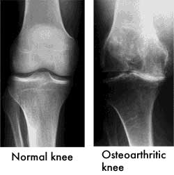
Knee Anatomy and Arthritis
Knee arthritis occurs when there is loss of cartilage of the knee. The knee joint has three bones: the femur (thigh bone), the tibia (shin bone), and the patella (kneecap). These three bones form knee compartments. The femur meets the tibia to form the joint, and the patella lies at the front of the patella. These three bones are covered with cartilage, which is a white polished material that allows for smooth gliding of the bones.
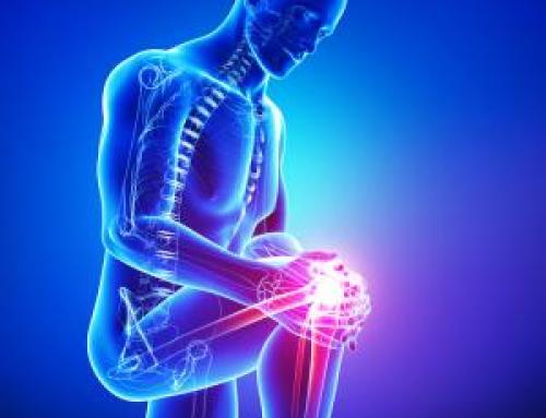
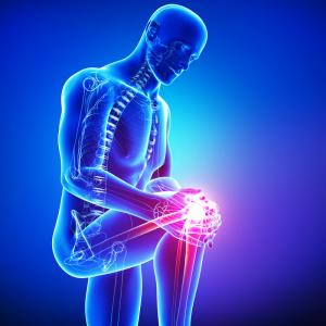
When osteoarthritis occurs, the knee joint is stiff and painful. Basically, bone rides against bone. The Beverly Hills orthopedic surgeon corrects this by realigning the angle made between the bones of the leg, so the patient can shift his or her body weight, allowing the healthy side to assume the majority of stress. Realigning the knee joint angle between the tibia and femur is called an osteotomy.
Benefits of High Tibial Osteotomy
 The high tibial osteotomy is helpful for patients with underlying pain and loss of range of motion from arthritis that affects one compartment of the knee. Once the load is decreased on the affected side of the knee, there is a delay in the need for a total joint replacement procedure. By reducing the heavy load, the pain and stiffness is also alleviated. In order to be a candidate for a high tibial osteotomy, you must have:
- Some ligament function around the knee
- Minimal arthritis in other compartments of the knee
- An adequate range of motion
If your orthopedic surgeon thinks you are a suitable candidate for this surgery, a high tibial osteotomy will reduce pain and improve overall knee function. The chance of the procedure offering pain relief five years following the procedure is around 80 percent. That percentage drops to 70 percent after 10 years and down to 50 percent after 15 years.
Before the Procedure
 Prior to undergoing a high tibial osteotomy, you will have x-rays and an MRI of the knee to assess and gage the degree of arthritis and structure damage. The x-rays show the doctor how the compartment is functioning, so he or she can predict where you are carrying the majority of your weight and how to properly realign the leg to achieve the best outcome. The MRI assesses the cartilage throughout all regions of the knee joint, as well as the knee ligaments and meniscus.
You are admitted to the hospital the day of surgery, and an anesthesiologist or anesthetist will discuss available options for anesthesia with you. You also are given prophylactic antibiotics to decreased risk of infection. When you are in the operating room, the anesthesia is given, and the surgical staff prepares your knee for the operation.
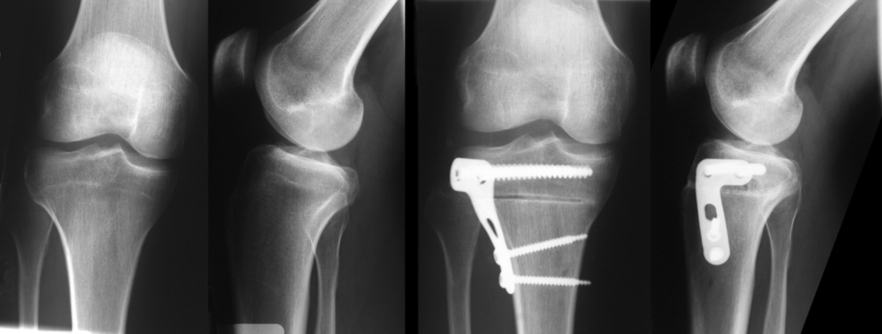
Two Methods of High Tibial Osteotomy
Closing wedge osteotomy – This procedure involves removing a piece of bone below the joint of the upper tibia. When arthritis has affected the medial knee compartment (as with varus knees), the bone wedge is taken from the outer region of the tibia. Once the bone segment is removed, the two bone ends are fixed together using a metal plate and pins.
Opening wedge osteotomy – In the wedge procedure, the orthopedic specialist cuts through the tibia on the inner side to open a wedge. This is often done by adding a segment of bone graft from the pelvic bone to hold the region open. The stabilize this, the surgeon inserts a plate and pins. This 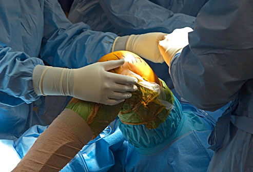 operation is most often done to the upper aspect of the tibia, right below the knee joint.
During the Procedure
During the high tibial osteotomy procedure, care is taken by the Los Angeles orthopedic surgeons to protect the blood vessels and nerves that are behind and around the knee joint. A small part of the fibula bone is used to in the closing wedge osteotomy, which is performed through the same incision.
Usually, a drain is inserted to allow the surgical wound to drain, and it will remain there for 24 hours. The incisions are closed and the leg is wrapped in a dry, sterile dressing. Many times, a brace will be required for a short time following the surgery.
After the Procedure
The patient is transported to a recovery room, where he or she is closely monitored following the high tibial osteotomy. An x-ray is taken of the knee to assess for alignment.
Once stable, the patient is taken to a hospital room and a physical therapist provides passive range of motion exercises. The day after surgery, you are taught to use crutches for up to three months. Most patients are able to go home three days after the procedure. Therapy continues once you are released to home, to help you improve mobility and regain muscular strength of the knee joint.
During the first week after the procedure, your leg will be swollen and the knee will feel stiff. Most orthopedic specialists prescribe regular pain medication during this time frame. It is vital for you to perform the exercises while at home to optimize recovery and rehabilitation. It may take up to six months to full recover from a high tibial osteotomy. Therefore, the most important aspect of rehabilitation is to strengthen the knee and allow it to heal.
Success Rate of a High Tibial Osteotomy
Around 70 percent of patients who have a high tibial osteotomy are satisfied with the results, and around 75 percent are able to return to sports and/or work activities. When the procedure fails, the orthopedic surgeon may suggest a total knee replacement. However, most patients who undergo this procedure have functioning knees at five to ten years following the surgery.
Risks of a High Tibial Osteotomy
All surgeries carry some risks and complications. The high tibial osteotomy is a safe procedure, but complications include:
- Bleeding
- Infection

- Blood clots of the legs
- Knee stiffness
- Damage to surrounding blood vessels and/or nerves
- Failure of the cut bone to heal
- Discomfort from metal-ware and subsequent removal
- Unexpected fractures
